- View all variations as list
| CODE | Title | Availability | Price | ||
|---|---|---|---|---|---|
|
|
ITM43762
|
In stock
|
Please sign in so that we can notify you about a reply
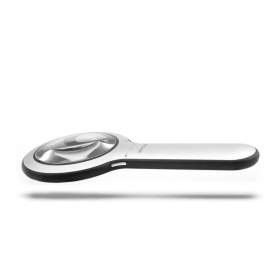
DermLite Lumio 2
Lumio 2 is the next generation in general skin exam illumination. By combining brightest-in-class polarized white light, three wavelengths in the UV spectrum, a special Wood mode, and special optical filters in a beautifully slim, quickly rechargeable device, Lumio 2 is the ultimate tool for visualizing various skin conditions.
Its large, 100mm diameter aspheric lens with 2.3x magnification allows for unrivaled visualization of dermatological structures in excellent optical clarity. Choose from polarized white light or one of three specific wavelengths: 365nm, 385nm in the UV spectrum, or 405nm blue light. Or, select its Wood mode, a special blend custom-tailored to resemble the unique illumination of a Wood's lamp which may help with evaluating the borders and extent of lentigo maligna as well as melanoma excision scars for signs of recurrence, among others. All five modes are available in three brightness levels.
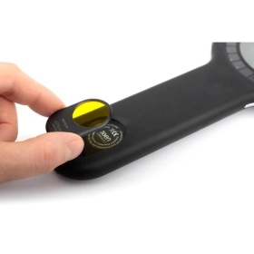
Lumio 2 comes with two magnetic OptiClip™ accessories. While one can be placed in the field of view to aid visualization, the other OptiClip may be stored in the handle.
- OptiClip 405nm Long-Pass – ideal for enhancing contrast of fluorescent features
- OptiClip 2.5x – boosts magnification by a factor of 2.5.
Lumio 2 integrates a powerful lithium-ion rechargeable battery designed to let you work for hours. Once its four-level charge indicator signals a low battery, recharging is fast and simple from any available USB outlet using the included USB-C to USB cable.
Model No. DLU2 is supplied with one Lumio 2, OptiClip 405nm Long-Pass, OptiClip 2.5x, and one protective neoprene pouch.
SNAPSHOT
- 100mm 2x lens
- ultra-bright white LEDs
- five lighting modes: white, 365nm, 385nm, 405mn, Wood
- three brightness levels
- lithium-ion rechargeable battery
- ultra-slim aluminum design with rubberized grip
- 4-level battery indicator
- OptiClip filter for enhanced fluorescence imaging
- OptiClip factor 2.5 magnification boost
- protective neoprene pouch
Warranty: 10 years
About the Wavelengths
365 nm and 385 nm are used mostly to map superficial pigmentation and high blood volume areas on the top 0.1 mm of the skin. In other words, these UV colors are largely absorbed in the epidermis, therefore pigmentation and superficial vascularity are highlighted by these UV frequencies. Pre-AK and BCC appear as darker areas, while lack of pigment like in vitiligo may show up brighter than the surrounding areas.
Having both 365 and 385 nm wavelengths enables differential visualization of disease interactive with higher vs lower frequency UV light. With both frequencies there is a fair amount of autofluorescence in tissue containing porphyrins.


